
Wound Healing — Physiology, Assessment, and Clinical Decision-Making
1. The four phases of normal wound healing
Wound healing is not four separate steps but a continuous, heavily overlapping biological response. Treat the phase concept as an interpretive framework: when a response lasts too long, is too weak, or fails to transition, it usually signals unresolved perfusion, infection, mechanical load, or host problems.
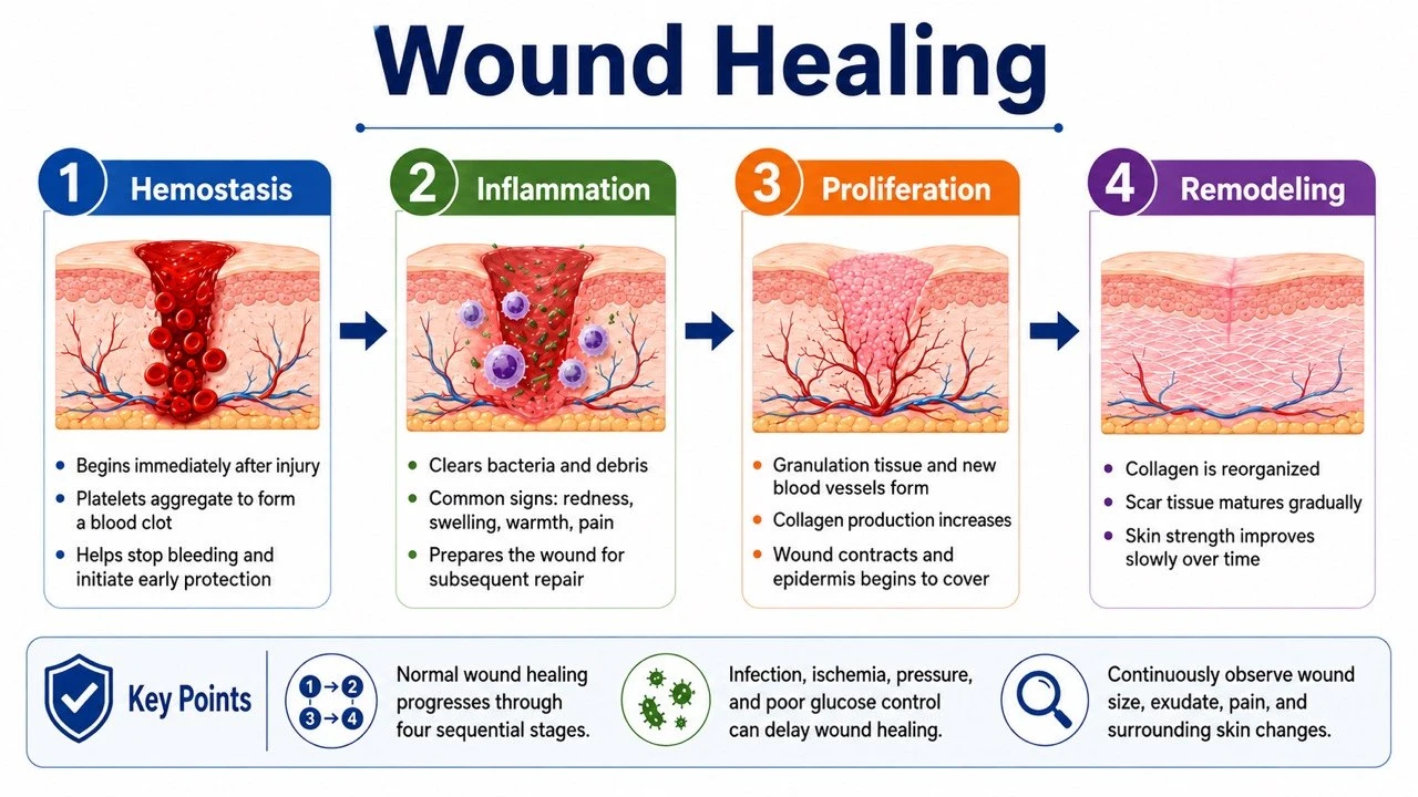
| Phase | Approximate timing* | Key cells / mediators | Clinical & tissue events |
|---|---|---|---|
| Hemostasis | Immediately to hours | Platelets, clotting factors, fibrin, PDGF, TGF-β | Vasoconstriction, platelet plug, fibrin network — a provisional matrix is built |
| Inflammation | Hours to ~day 3–5 | Neutrophils, macrophages, cytokines, MMPs | Clearing pathogens and necrotic tissue; macrophage phenotype switching helps the transition into proliferation |
| Proliferation | ~Day 3 to weeks | Fibroblasts, keratinocytes, endothelial cells | Angiogenesis, granulation, collagen deposition, contraction, re-epithelialization |
| Maturation / Remodeling | Weeks to months+ | Fibroblasts, myofibroblasts, MMP/TIMP balance | Collagen III→I conversion, cross-linking and realignment; tensile strength increases but rarely returns to uninjured skin |
*Times are for orientation only; phases overlap and depend on wound depth, perfusion, infection, and host status.
2. Healing patterns and wound closure
Closure choice depends on contamination, tissue loss, perfusion, tension, and infection risk — not on the speed of suturing. If the cause is uncontrolled or necrosis and dead space persist, premature closure can seal infection inside; conversely, proper approximation shortens the repair distance and reduces scarring.
| Pattern | Features | Clinical points |
|---|---|---|
| Primary intention | Clean incision, approximated edges, minimal tissue loss | Less granulation and scarring; still monitor for hematoma, infection, dehiscence |
| Secondary intention | Open defect closing by granulation, contraction, epithelialization | Slower, more scarring; manage dead space, exudate, pressure and etiology |
| Tertiary / delayed primary closure | Higher contamination or infection risk — treat open first, close later | Re-evaluate closure timing once contamination, infection and necrosis are controlled |
3. Biological differences: acute vs. chronic wounds
Acute wounds progress predictably from inflammation to proliferation and remodeling; chronic wounds are trapped in an unbalanced inflammatory environment. This explains why changing dressings alone is usually insufficient: with persistent ischemia, repetitive pressure, biofilm, or systemic metabolic problems, newly formed tissue keeps being damaged.
| Aspect | Acute healing wound | Chronic / stalled wound |
|---|---|---|
| Inflammation | Starts and resolves on schedule | Persistent inflammation, unbalanced macrophage switching |
| Proteases | MMPs and inhibitors balanced | Elevated protease activity; growth factors and ECM degraded |
| Bacterial load | Host controls contamination | Possible biofilm, recurrent or deep infection |
| Cell response | Normal fibroblast/keratinocyte migration & proliferation | Cell senescence, poor migration, stalled edge epithelium |
| Perfusion / mechanics | Oxygenation and mechanics support repair | Persistent ischemia, pressure, shear, edema, or repeated trauma |
4. Local and systemic factors affecting healing
The wound surface is the product of an interaction between systemic status and the local environment. Assessment should review "locally treatable factors" and "systemic limiting conditions" side by side, distinguishing what can be corrected immediately, what needs cross-specialty input, and which factors can only be risk-reduced rather than fully reversed.
| Category | Common factors | Assessment / management direction |
|---|---|---|
| Perfusion & oxygenation | PAD, venous hypertension, edema, hypoxia, anemia | Exam plus vascular testing; revascularization, compression, or edema control by etiology |
| Infection & inflammation | Necrosis, abscess, biofilm, foreign body, osteomyelitis | Debridement, drainage, proper specimens and anti-infective therapy; do not rely on surface swabs |
| Mechanical | Pressure, shear, friction, repetitive loading or tension | Offloading, turning, fixation and protection; correct the injuring force |
| Moisture balance | Dryness, excess exudate, maceration, incontinence damage | Choose dressings that keep moisture balanced; protect periwound skin |
| Metabolic & nutrition | Diabetes, renal disease, low protein intake, weight loss | Metabolic control; risk-based nutrition assessment and individualized supplementation |
| Drugs & lifestyle | Steroids, immunosuppressants, chemotherapy, smoking, alcohol | Review modifiable factors; smoking cessation and multidisciplinary coordination |
| Atypical causes | Vasculitis, pyoderma gangrenosum, malignancy, atypical infection | If lesions look atypical or worsen after debridement — dermatology, pathology and cultures |
5. Clinical assessment: judge healability first, then prepare the wound bed
The core of assessment is not choosing a dressing first but answering three questions: is the etiology clear, is perfusion adequate, and is the current goal healing, maintenance, or symptom palliation? For the ischemic limb in particular, the safety of debridement and compression must rest on vascular assessment.
- Etiology & reversibility: wound type, duration, mechanism, prior treatment, recurrence pattern
- Perfusion: skin temperature, color, pulses, capillary refill; ABI, TBI, toe pressure, TcPO₂ or imaging when needed
- Infection: local inflammation, pus, odor, abscess, necrosis, systemic response; classify diabetic foot infection clinically, not by culture alone
- Tissue: granulation, slough, eschar, tendon/bone exposure, undermining, sinus tracts, dead space
- Moisture: exudate volume, viscosity, color, dressing saturation, periwound maceration
- Edge & periwound: epithelial advance, rolled edges, callus, dermatitis, edema, induration, pain
- Host factors: glycemia, nutrition, renal function, immunity, drugs, mobility, support system, care capacity
6. Quantitative tracking and interpreting response
A single snapshot only describes the moment; only serial, standardized data can reveal a trend. Size, tissue, exudate, pain and function should be recorded with consistent methods. Photos strengthen comparison but must still be combined with palpation, depth, undermining, perfusion and infection signs — do not mistake image change for the full clinical picture.
| Indicator | What to record | Interpretation cautions |
|---|---|---|
| Size | Length, width, depth; area/volume when needed | Use a consistent method; L×W may overestimate irregular wounds |
| Tissue proportions | Epithelium, granulation, slough, eschar | Observer-dependent; pair with photos and trends |
| Exudate & odor | Amount, color, viscosity, odor, dressing wear time | Odor and purulence must be read with clinical signs of infection |
| Pain & function | Rest / dressing-change / weight-bearing pain, sleep, activity | Sudden pain increase may mean infection, ischemia, or unrelieved pressure |
| Imaging (photos) | Fixed distance, angle, lighting, scale, date | Consent and privacy; photos never replace palpation and depth assessment |
| Healing velocity | Percent area reduction, epithelial advance | For many DFUs, <50% area reduction at 4 weeks triggers reassessment — do not apply mechanically to all wounds |
7. Core measures that support healing
Treatment strategy has four layers: etiologic correction, wound-bed preparation, systemic support, and behavioral execution. Dressings mainly protect tissue and regulate moisture; what actually determines sustained healing is usually perfusion, offloading, infection control, metabolic and nutritional status, and whether the patient can carry out the care plan.
- Treat the cause: revascularization, venous/lymphedema control, offloading, pressure redistribution, releasing sustained tension
- Debridement & infection control: choose method by perfusion, tissue, pain and goals; drain abscesses; evaluate deep infection
- Moisture balance: choose dressings by exudate; avoid desiccation, maceration and unnecessary frequent changes (wet gauze principles)
- Protect new tissue: low-trauma contact layers; avoid routine wet-to-dry gauze that destroys granulation and epithelium
- Metabolism & nutrition: correct hyperglycemia, energy and protein deficits; treat dehydration, anemia, micronutrient deficiencies
- Patient engagement: pain control, smoking cessation, education, shared goals, and an executable follow-up plan
8. Stalled and abnormal healing
A stall should not be summarized as merely "healing slowly" — it calls for re-examining the diagnosis and treatment assumptions. If a previously improving wound suddenly deteriorates, enlarges rapidly after debridement, shows pain out of proportion to appearance, or develops abnormal bleeding or a mass, prioritize excluding ischemia, deep infection, inflammatory disease, and malignancy.
| Situation | Possible clues | Reassess first |
|---|---|---|
| Chronic inflammation / stall | No size change, recurrent slough, dusky granulation, non-advancing edge | Etiology, perfusion, infection/biofilm, pressure, dressing strategy, adherence |
| Hypergranulation | Granulation above skin level, bleeds easily, blocks epithelium | Friction, excess moisture, foreign body, infection, local irritation |
| Dehiscence | Incision separation, rising exudate, deep tissue exposure | Infection, hematoma, tension, nutrition, fascial integrity; deep dehiscence is urgent |
| Hypertrophic scar | Confined to wound, red firm raised, itchy/painful | Tension, depth, site, personal risk; early silicone, pressure, or specialist care |
| Keloid | Extends beyond original wound, keeps growing | History and high-risk sites; avoid ill-advised simple excision |
| Abnormal bleeding / mass | Friable, everted, rapidly growing, non-healing | Exclude malignancy, vascular lesions or atypical infection; consider biopsy |
9. Signals for escalation or referral
The purpose of referral is not to hand off dressing changes but to obtain diagnostics or treatment beyond the current level of care — vascular intervention, imaging and bone-infection workup, extensive debridement, reconstructive surgery, pathologic diagnosis, or systemic immunotherapy. Limb- or life-threatening situations take emergency priority.
- Suspected acute limb ischemia, rapidly progressing necrosis, necrotizing soft-tissue infection, or sepsis
- Deep abscess, osteomyelitis, exposed vessels/implants, or fascial dehiscence
- Diabetic foot with moderate-to-severe infection, ischemia, Charcot suspicion, or failed offloading
- No reasonable improvement despite appropriate standard care, or uncertain diagnosis
- Rapid enlargement after debridement, violaceous undermined edges, or pain out of proportion — consider inflammatory/vasculitic wounds
- Recurrent bleeding, everted mass, or long-standing non-healing — exclude malignant wounds
10. Minimum documentation dataset
Documentation should let the next caregiver reconstruct the wound's etiology, current state, interventions taken, and the thresholds for subsequent decisions. Beyond size, explicitly record the measurement method, perfusion and infection judgments, how offloading or compression is being carried out, and which changes should trigger an earlier visit or escalation of care.
| Field | Minimum content |
|---|---|
| Basics | De-identified ID, date, site, etiology/diagnosis, wound duration |
| Objective measures | L/W/D, undermining/sinus, tissue proportions, exudate, odor, periwound skin |
| Patient status | Pain, perfusion, infection, glycemia/nutrition, mobility and pressure exposure |
| Interventions | Cleansing, debridement, dressing, offloading/compression, drugs, education, referral |
| Follow-up plan | Dressing frequency, reassessment date, deterioration triggers, contact/escalation route |
Last updated: 2026-09-01 ・ Patient version: How do wounds heal? (Chinese)